Page 72 - 《渔业研究》2025年第5期
P. 72

第 5 期              孔维光等: Ⅱ型草鱼呼肠孤病毒           VP4  兔源单克隆抗体的制备和鉴定                        613

              的病毒    VP4  抗原特异性荧光信号;而在健康对照                     (图  4) 。以上结果表明,1D5、1F9           和  1H1  细胞
              组中未检测到阳性荧光信号,证实了这些单克隆抗                           株上清液具有良好的抗原特异性,可用于重组单克
              体对   GCRV-Ⅱ病毒抗原有较强的特异性识别能力                       隆抗体制备的候选细胞株。
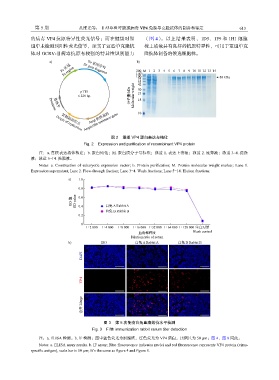
                      a)               S6 基因序列             b)
                           Fc 区域     S6 gene fragment      200 M 1 2 3 4 5 6 7 8 9 10 11 12 13 14
                            Fc region
                                                           140
                                                           100                                 80 kDa
                                                            75
                                                            55
                                                            42
                                   pTT5                     30
                                                            23
                                  6 229 bp              分子量/kDa  Molecular weight  18
                        启动子
                      Promoter
                                       Amp 抗性基因
                         Origin of replication Ampicillin resistance gene  10
                           复制起始位点

                                                图 2    重组  VP4  蛋白表达与纯化
                                     Fig. 2    Expression and purification of recombinant VP4 protein
                  注:a. 真核表达载体构建;b. 蛋白纯化;M. 蛋白质分子量标准;泳道                 1. 表达上清液;泳道     2. 流穿液;泳道    3~4. 洗涤
              液;泳道   5~14. 洗脱液。
                  Notes:  a.  Construction  of  eukaryotic  expression  vector;  b.  Protein  purification;  M.  Protein  molecular  weight  marker;  Lane  1.
              Expression supernatant; Lane 2. Flow-through fraction; Lane 3−4. Wash fractions; Lane 5−14. Elution fractions.

                              a)   1.0
                                   0.8
                                OD 值  OD value  0.6  白兔 A Rabbit A
                                   0.4
                                              白兔 B Rabbit B
                                   0.2
                                    0
                                      1∶2 000  1∶4 000  1∶8 000 1∶16 000 1∶32 000 1∶64 000 1∶128 000 空白对照
                                                            血清稀释度                  Blank control
                                                         Dilution ratio of serum
                              b)            ISO            白兔 A Rabbit A     白兔 B Rabbit B

                                    DAPI


                                    VP4


                                    合并 Merge


                                             图 3    第  5  次免疫白兔血清效价水平检测
                                        Fig. 3    Fifth immunization rabbit serum titer detection

                  注:a. ELISA  检测。b. IF  检测;图中蓝色荧光为细胞核,红色荧光为          VP4  蛋白,比例尺为    50 µm;图  4、图  8  同此。
                  Notes: a. ELISA assay results. b. IF assay; Blue fluorescence indicates nuclei and red fluorescence represents VP4 protein (virus-
              specific antigen), scale bar is 50 µm; It’s the same as figure 4 and figure 8.
   67   68   69   70   71   72   73   74   75   76   77