Page 46 - 《水产学报》2026年第01期
P. 46

1 期                                     水    产    学    报                                 50 卷


                                                                      H. sapiens PDB:1f45 M. musculus PDB:8cr6
                                                                            p35
                                                                                       p35  M. musculus
                                                                           p40      p40
                                                                                                 180°
                                                                                        H. sapiens  M. salmoides

                                                                                          p35  p35  p35a
                                                   p35a+p40a
                     M. musculus PDB: 8cr6
                            (a)                         (b)             (c)       (d)          (e)
                                                                     M. salmoides  M. salmoides  M. salmoides
                                                                              p35a      p35a      p35a

                                                                            p40a                 p40c
                                                                                      p40b


                         p35a+p40b                 p35a+p40c

                            (f)                         (g)              (h)        (i)         (j)
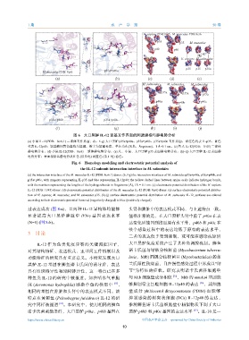
                                    图 6    大口黑鲈  IL-12  亚基互作界面的同源建模与静电势分析
              (a) 小鼠  IL-12(PDB:8cr6.1) 二聚体互作界面;(b,f~g) 大口黑鲈  p35a+p40a、p35a+p40b、p35a+p40c 互作界面,洋红色代表  IL-p35,蓝色
              代表  IL-12p40;氨基酸间黄色虚线为氢键,数字为氢键长度,单位为Å (埃米,Ångstrom),1 Å=0.1 nm。(c) 智人       IL-12(PDB:1F45) 二聚体
              静电势分布。(d) 小鼠   IL-12(PDB:8cr6) 二聚体静电势分布。(e) 人、小鼠、大口黑鲈     p35  表面静电势分布。(h)~(j) 大口黑鲈  IL-12  表面静
              电势分布;表面根据其静电势从红色         (带负电) 到蓝色  (带正电) 着色。
                                   Fig. 6 Homology modeling and electrostatic potential analysis of
                                        the IL-12 subunit interaction interface in M. salmoides
              (a) the interaction interface of the M. musculus IL-12 (PDB: 8cr6.1) dimer. (b, f-g) the interaction interfaces of M. salmoides p35a+p40a, p35a+p40b, and
              p35a+p40c, with magenta representing IL-p35 and blue representing IL-12p40; the yellow dashed lines between amino acids indicate hydrogen bonds,
              with the numbers representing the lengths of the hydrogen bonds in Ångstroms (Å), 1Å = 0.1 nm. (c) electrostatic potential distribution of the H. sapiens
              IL-12 (PDB: 1F45) dimer. (d) electrostatic potential distribution of the M. musculus IL-12 (PDB: 8cr6) dimer. (e) surface electrostatic potential distribu-
              tion of H. sapiens, M. musculus, and M. salmoides p35. (h)-(j) surface electrostatic potential distribution of M. salmoides IL-12; surfaces are colored
              according to their electrostatic potential from red (negatively charged) to blue (positively charged).
              过表达成功      (图  8-a),且两种    IL-12  异构体均能够         头肾和脾脏中的表达模式不同,与上述特点一致。
              显著提高大口黑鲈脾脏中               IFN-γ 基因表达水平           值得注意的是,在大口黑鲈头肾中除了                  p40a  在表
              (N=5) (图  8-b)。                                  达变化后能回到接近原有水平外,p40b                和  p40c 在

                                                               整个感染过程中的表达均低于原有的表达水平,
               3    讨论
                                                               二者的表达似乎受到抑制,或可能在感染病原后
                   IL-12  作为鱼类免疫应答的关键调控因子,                     大口黑鲈免疫系统产生了某种负调控机制。鰤鱼
              对其结构特征、表达模式、亚基间互作机制以及                            诺卡氏菌与结核分枝杆菌            (Mycobacterium tubercu-
              功能特征的研究具有重要意义。本研究发现大口                            losis,Mtb) 同属分枝杆菌目       (Mycobacteriales) 的革
              黑鲈   IL-12  基因在鰤鱼诺卡氏菌的诱导后,表达                     兰氏阳性致病菌,且在慢性感染过程中形成以“结
              具有组织特异性和时间特异性,这一特点已在多                            节”为特征的症状。研究表明诺卡氏菌科细胞壁
                                                                                    [35]
              种鱼类    IL-12  的研究中被报道。如在嗜水气单胞                    与  Mtb  细胞壁成分相似  ,Mtb       的  mmAa4  基因能
                                                                                                 [36]
              菌  (Aeromonas hydrophila) 感染草鱼的模型中        [33] ,  够抑制宿主巨噬细胞         IL-12p40  的表达  ,其细胞
              相同的基因在脾脏和头肾中的表达模式不同。该                            壁成分    phthiocerol dimycoserosate (PDIM) 也能够
              特点在黄颡鱼        (Pelteobagrus fulvidraco) IL-12  的研  抑制感染的树突状细胞          (DCs) IL-12p40  的表达,
              究中同样被报道         [34] 。本研究中,使用相同的鰤鱼               推测鰤鱼诺卡氏菌细胞壁中相似物质下调了大口
              诺卡氏菌刺激条件,大口黑鲈               p40a、p40b  基因在       黑鲈  p40b  和  p40c 基因的表达水平  。IL-10       是一
                                                                                              [37]
              https://www.china-fishery.cn                           中国水产学会主办    sponsored by China Society of Fisheries
                                                            10
   41   42   43   44   45   46   47   48   49   50   51