Page 68 - 《水产学报》2025年第11期
P. 68

苏金枝,等                                                                水产学报, 2025, 49(11): 119606

              的脂滴。对照组与           AST800  组细胞核直径和面              (P<0.05) (表  2)。相比对照组,AST800         组肝细
              积无显著性差异         (P>0.05),AST800   组肝细胞的          胞内自噬小体、自噬溶酶体和线粒体数量均显
              直径和面积相比对照组显著减小                  (P<0.05),细       著增多    (P<0.05),脂滴数量显著减少            (P<0.05)
              胞 内 脂 滴 直 径 和 面 积 相 比 对 照 组 显 著 减 小              (表  3)。

                                      表 2    对照组和  AST800  组赤点石斑鱼肝细胞结构量化表
                      Tab. 2    Quantitative table of hepatocyte structure of E. akaara in control group and AST800 group
                      组别         细胞核直径/µm 细胞核面积/µm     2   脂滴直径/µm     脂滴面积/µm  2  肝细胞直径/µm     肝细胞面积/µm 2
                      group      nuclear diameter  nuclear area  lipid droplet diameter lipid droplet area hepatocyte diameter  hepatocyte area
               对照组 control group   5.69±0.98   26.53±4.16   1.81±0.65 a  2.82±0.51 a  28.89±2.86 a  718.52±65.23 a
               AST800组 AST800 group  6.08±0.84  28.18±6.96  0.98±0.15 b  1.06±0.19 b  17.44±2.34 b  276.00±42.67 b
              注:同列数据肩标不同小写字母表示差异显著(P<0.05),下同。
              Notes: In the same column, values with different small letter superscripts mean significant difference (P<0.05), the same below.


                                    表 3    对照组和  AST800  组赤点石斑鱼单个肝细胞中自噬水平
                          Tab. 3    Autophagy level in hepatocytes of E. akaara in control group and AST800 group
                       组别               自噬小体数量            自噬溶酶体数量              脂滴数量            线粒体数量
                       group         autophagosome number  autophagolysosome number  lipid droplet number  mitochondria number
                对照组 control group       49.67±7.74 b        28.10±5.32 b      65.80±11.54 a   506.11±84.20 b
                AST800组 AST800 group    86.17±15.37 a       61.90±17.51 a     15.10±3.61 b    669.78±121.89 a
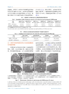
               2.3    饲料中添加虾青素对赤点石斑鱼肠道结构                       整齐的刷状缘;上皮细胞之间紧密连接和黏附

              和自噬的影响                                           连接结构清晰、完整           (图版Ⅱ)。其中对照组肠
                   对照组和     AST800  组柱状肠上皮细胞排列                 上皮细胞游离端可见部分线粒体有空泡、嵴较
              整齐,面积、形态无明显差别,微绒毛排列规                             少的现象。AST800       组细胞内线粒体呈椭圆形和
              则,其细胞膜清晰完整,在上皮细胞顶端形成                             长条形,相比对照组细胞质中线粒体嵴完整,


                                                                         ·
                                           TJ                                                     TJ
                                                     M                                             AJ
                                         MV AJ                    
                                                                       ¶              MV
                                                                         ¶  ·
                              CC                                 ·     ·
                             N  GC               GC                       ¶ ·
                                   MV                               ¶     
                               RER                                            
                                                                     LD 
                    5 μm   BL         1 1  1 μm           2 2  0.2 μm          3 3  0.5 μm         4 4
                                                                          ¶  
                                            MV                     ·
                                                                · M ·
                                   MV           TJ           ¶      ¶  ·  ·             MV
                         GC                    AJ     GC       ·  ·      ¶  
                                                                 
                     CC                                            ·  ·  
                                           M                     ·     
                     N    RER                                            
                                                                      
                    5 μm              5  2 μm             6  0.2 μm ·  ¶  ·    7  0.5 μm        M  8
                                       5                   6      ·             7                   8
                               图版 Ⅱ    透射电镜观察对照组和         AST800  组赤点石斑鱼肠道组织超微结构
              1. 对照组肠道黏膜层上皮细胞结构;2. 正在分泌黏液的杯状细胞;3. 柱状上皮细胞游离面结构;4. 柱状上皮细胞微绒毛状结构;5.
              AST800  组肠道黏膜层上皮细胞结构;6. 正在分泌黏液的杯状细胞;7. 柱状上皮细胞游离面结构;8. 柱状上皮细胞微绒毛状结构。 GC. 杯
              状细胞; CC. 柱状细胞; BL. 基底面;TJ. 紧密连接;AJ.黏附连接。
                            Plate Ⅱ Ultrastructure of intestine in control group and AST800 group of E. akaara
              1. epithelial cell structure of intestinal mucosa in control group; 2. goblet cell secreting mucus; 3. free surface structure of columnar epithelial cell;
              4. microvilli structure of columnar epithelial cell; 5. epithelial cell structure of intestinal mucosa in AST800; 6. goblet cell secreting mucus; 7. free sur-
              face structure of columnar epithelial cell; 8. microvilli structure of columnar epithelial cell. GC. goblet cell; CC. columnar cell; BL. basal surface; TJ.
              tight junction; AJ. adherens Junction.

              中国水产学会主办  sponsored by China Society of Fisheries                          https://www.china-fishery.cn
                                                            5
   63   64   65   66   67   68   69   70   71   72   73