Page 67 - 《水产学报》2025年第11期
P. 67

苏金枝,等                                                                水产学报, 2025, 49(11): 119606

              型和二次多项式模型均显示随着虾青素添加量                                   4.0      线性模型`linear model
                                                                     3.8
              的增加,赤点石斑鱼的肝脏               HSI 呈现下降趋势,                 3.6      二次多项式模型`second order
                                                                              polynomial model
                                                         2
              线性回归模型:          Y HSI = −0.000 54x+2.740 49(R =       3.4
                                                        2
              0.949 91);二次多项式模型:           Y HSI = 0.000 44x −       3.2   a  ab  ab     y=−0.000 54x+2.740 49
                                                                     3.0
                                 2
                                                                                             2
              0.008 99x+2.773 78(R =0.979 63),二次多项式回             肝体指数/%  hepatosomatic index  2.8  ab  R =0.949 91  b
              归方程拟合效果优于线性回归方程                  (图  1)。               2.6
                                                                     2.4
               2.2    饲料中添加虾青素对赤点石斑鱼肝脏结构                             2.2
                                                                     2.0        y=0.000 44x −0.008 99x+2.773 78
                                                                                        2
                                                                                        2
              和自噬的影响                                                 1.8               R =0.979 63
                                                                     1.6
                   对照组和     AST800  组肝细胞的细胞核内染                        −100 0 100 200 300 400 500 600 700 800 900
              色质分布均匀,常染色质较多,异染色质较少,                                             虾青素添加量/(mg/kg)
              核孔清晰,可见         0~2  个核仁   (图版Ⅰ);细胞质                           astaxanthin supplemental level
              中均有少量自噬小体,周围环绕粗面内质网和                                图 1    饲料中添加不同浓度虾青素的赤点石斑鱼
                                                                             HSI 回归分析模型
              溶酶体,并出现脂滴分解现象,可见分割状边
                                                               图中不同上标字母表示差异显著        (P<0.05)。
              缘;内皮细胞和肝细胞之间窦周隙明显,窦周
                                                                 Fig. 1 Regression analysis model of E. akaara with
              隙结构无显著性差异。其中对照组细胞质内分
                                                                   different concentrations of astaxanthin in feed
              布不均匀,细胞核和细胞器偏向一边,脂滴较
                                                               Different superscript letters in the figure indicate significant differences
              大且形状规则;AST800          组细胞质内分布均匀,                (P<0.05).
              脂滴自噬小体中明显可见内容物为形状不规则


                                              ¶   NP  N
                                         ¶     ¶                     RBC
                       Hep                         NE  ¶                               DS        RBC
                                               ·  ¶  RER  SER            CV              NETC
                       GL                        ¶    ·                     FSC    M
                        SER     LD              ¶                EN
                    RER    N
                               M                M Ly
                                                  ·
                                Ly   KC   · ¶  LD                            Hep
                    5 μm              1  1 μm             2  5 μm              3  1 μm             4
                                  MV
                                                                                       RBC
                                                GL
                    LD  Hep                   
                                                                     RBC                      DS
                                                 SER                                      NETC
                          M                           ·
                                     MV           ·     ·
                             N            · RER                           EN
                          Ly         BC   ¶       NE       ·            HS
                                         Ly  ·
                    5 μm     LD       5  1 μm  NP NU  N   6  2 μm              7  1 μm       MV    8
                                                                                      FSC
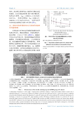
                               图版 Ⅰ    透射电镜观察对照组和         AST800  组赤点石斑鱼肝脏组织超微结构
              1. 对照组肝细胞超微结构;2. 肝细胞局部放大结构;3. 以中央静脉为中心的肝板结构;4. 内皮细胞结构;5. AST800                组肝细胞超微结构;
              6. 肝细胞局部放大结构;7. 肝血窦结构;8. 肝血窦内壁上的内皮细胞。Hep. 肝细胞;N.细胞核;NP. 核孔;NE. 核膜;NU. 核仁;M. 线粒
              体;LD. 脂滴;Ly. 溶酶体;GL. 糖原;RER. 粗面内质网;SER. 滑面内质网;CV. 中央静脉;BC. 胆小管;HS. 肝血窦;DS. 窦周隙;KC.
              枯否细胞;EN. 内皮细胞;NETC. 内皮细胞核;FSC. 肝星状细胞;MV. 微绒毛;RBC. 红细胞;实心三角形              (▲) 表示形态正常、嵴较多的
              线粒体;空心三角形      (△) 表示出现肿胀、空泡化的线粒体;黑色单箭头          (→) 表示自噬小体   (脂滴自噬小体、线粒体自噬小体);黑色双箭
              头表示自噬溶酶体,下同。
                            Plate Ⅰ Ultrastructure of liver in the control group and AST800 group of E. akaara
              1.  ultrastructure  of  hepatocyte  in  control  group;  2.  hepatocyte  local  amplification  structure;  3.  hepatic  plate  structure  centered  on  central  vein;  4.
              endothelial cell structure.; 5. ultrastructure of hepatocyte in AST800; 6. hepatocyte local amplification structure; 7. hepatic sinus structure; 8. endothelial
              cell on the wall of the hepatic sinusoid. Hep. hepatocyte; N. nucleus; NP. nuclear pore; NE. nuclear membrane; NU. nucleolus; M. mitochondria; LD.
              lipid droplet; Ly. lysosome; GL. glycogen; RER. rough endoplasmic reticulum; SER. slippery endoplasmic reticulum; CV. central vein; BC. bile can-
              aliculus; HS. hepatic sinusoid; DS. Disse's space; KC. Kupffer cell; EN. endothelial cell; NETC. endothelial cell nucleus; FSC. hepatic stellate cell; MV.
              microvilli; RBC. red blood cell; the solid triangle (▲) indicates the mitochondria with normal form and more mitochondrial cristae; the hollow triangle
              (△) indicates swollen, vacuolated mitochondria; the black single arrow ( →) represents autophagosome (lipid droplet autophagosome, mitochondrial
              autophagosome); the black double arrow indicates the autophagolysosome, the same below.

              https://www.china-fishery.cn                           中国水产学会主办    sponsored by China Society of Fisheries
                                                            4
   62   63   64   65   66   67   68   69   70   71   72