Page 144 - 《中国药科大学学报》2026年第2期
P. 144

270                      学报   Journal of China Pharmaceutical University 2026, 57(2): 266 − 274  第 57 卷

                3.2    hUCMSCs 的成脂、成骨、成软骨分化能力                   潜力   (图  2-A);茜素红染色显示       hUCMSC   的成骨分
               结果                                               化 潜 力   (图  2-B); 阿 尔 辛 蓝 染 色 显 示   hUCMSCs
                    油红   O  染色显示    hUCMSCs 的脂肪发生分化             的软骨分化潜力        (图  2-C)。

                  A                               B                             C


                                       100 μm                          100 μm                         100 μm

               Figure 2    Results of adipogenic (A), osteogenic (B) and cartilage (C) differentiation abilities of hUCMSCs

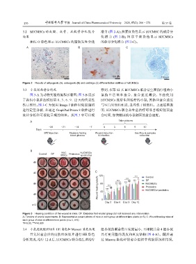
                3.3    小鼠创面愈合情况                                 察到,在第     12 天  hUCMSCs 联合益生菌治疗组的小
                    图  3-A  为动物实验的流程示意图;图            3-B  展示    鼠 伤 口 已 基 本 愈 合 , 愈 合 速 度 最 快 , 单 独 使 用
               了各组小鼠在造模后第            0、3、6、9、12 天的代表性           hUCMSCs 或益生菌治疗的小鼠,其伤口愈合速度
               伤口照片;图       3-C  为使用  Image J 软件对创面面积           与空白对照组相近,且均快于模型组。上述结果表
               进行定量分析,并通过           GraphPad Prism 8 软件进行       明,hUCMSCs 联合益生菌治疗可显著缩短创面愈
               统计分析和可视化呈现的结果。从图                   3 中可以观        合时间,加快糖尿病小鼠创面的愈合速度。

                                                                           Take pictures
                    A
                                −26    −21      −14      −7       0     3      6     9     12
                        Days
                             STZ injection  Measure fasting   Wound induction        Sacrifice & samples
                                             blood glucose    & infection               collection


                    B
                                        hUC
                         Control  DF   MSCs  Probiotics  hUCMSCs
                                                   +probiotics
                      d0

                                                            C
                      d3
                                                               120              ***  ***  *  Control
                                                              Wound healing rate/%  ***  ns  Probiotics
                                                                          ***
                      d6                                        80  ***  *** ns  ***  ns  ***  DF
                                                                                            hUCMSCs
                                                                                            hUCMSCs+probiotics
                      d9                                        40
                                                                0
                                                                   Day 3  Day 6  Day 9 Day 12
                     d12


               Figure 3    Healing condition of the wound in mice. DF: Diabetes foot-model group did not received any intervention
               A: Timeline of animal experiments; B: Representative wound photos of mice in each group at different time points (n=5); C: Wound healing rates of
               each group of mice at different time points ( x ± s, n=5)
               *P<0.05; ***P<0.001

                3.4    小鼠皮肤组织切片     HE  染色和   Masson 染色结果       组小鼠的剩余伤口宽度最小,且相较其余                   4 组小鼠
                    首先对愈合后的皮肤组织切片进行                  HE  染色     具有更完整的表皮和真皮结构(图                 4-A)。随后通
               分析发现,治疗       12 d 后,hUCMSCs 联合益生菌治疗             过  Masson 染色评估愈合组织中的胶原沉积情况,
   139   140   141   142   143   144   145   146   147   148   149