
Page 47 - 《中国药科大学学报》2025年第5期
P. 47
第 56 卷第 5 期 齐 磊,等:二甲双胍通过促进醛酮还原酶 AKR1C3 降解抑制肝细胞癌恶性进展的机制研究 579
A HepG2 HepG2 HCC-LM3 HCC-LM3
CHX + + + + CHX + + + + CHX + + + + CHX + + + +
Metformin − + − + Metformin − + − + Metformin − + − + Metformin − + − +
Chloroquine − − + + MG132 − − + + Chloroquine − − + + MG132 − − + +
AKR1C3 AKR1C3 AKR1C3 AKR1C3
β-actin β-actin β-actin β-actin
250 ** 150 n.s. 200 ** 150
Relative AKR1C3 level /(% of control) 200 Relative AKR1C3 level /(% of control) 100 Relative AKR1C3 level /(% of control) 100 Relative AKR1C3 level /(% of control) 100 n.s.
150
150
100
50
50
50
50
CHX 0 + + + + CHX 0 + + + + CHX 0 + + + + CHX 0 + + + +
Metformin − + − + Metformin − + − + Metformin − + − + Metformin − + − +
Chloroquine − − + + MG132 − − + + Chloroquine − − + + MG132 − − + +
B C D
Metformin HBSS starvation c(chloroquine)/(mmol/L)
t/h 0 24 36 48 t/h 0 2 4 150 0 10 20 40 200
LC3-I AKR1C3 ** AKR1C3 **
LC3-II SF 100 n.s. 150 n.s.
LC3-I LF LC3-I LC3-I n.s.
LC3-II LC3-II Relative AKR1C3 level /(% of control) LC3-II Relative AKR1C3 level /(% of control) 100
p62 p62 50 p62 50
β-actin β-actin β-actin
HCC-LM3 0 0 0 10 20 40
HCC-LM3 0 2 4
t/h c(chloroquine)/(mmol/L)
E Metformin − − − + + + − − − 120 F
Relative AKR1C3 level /(% of control) 80 PBS ** ** AKR1C3
Chloroquine − − − − − − + + + Input IP:LC3
HBSS starvation 0 2 4 0 2 4 0 2 4 100 Chloroquine + + + + − + +
Metformin −
LC3-I
LC3-II
Metformin
2
1
HCC-LM3 60 0 Chloroquine 3 4 5 β-actin HCC-LM3
t/h
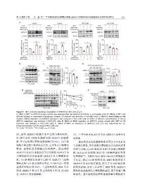
Figure 4 MET promotes autophagic degradation of AKR1C3 in HCC cells ( x ± s)
A: Effect of MET on AKR1C3 protein content was detected after the treatment of MG132 or chloroquine (CQ); B: Effects of MET with
different duration on expression of autophagic markers LC3 and p62 was detected in HCC-LM3 cells; C: Effect of Hank's Balanced Salt
Solution (HBSS) starvation on AKR1C3 expression was analyzed in HCC-LM3 cells; D: Effect of different concentrations of CQ on
AKR1C3 expression was detected in HCC-LM3 cells; E: Effect of HBSS starvation on AKR1C3 protein content after MET or CQ
treatment was detected in HCC-LM3 cells; F: Effect of MET on interaction of AKR1C3 with LC3 was determined by Co-
Immunoprecipitation (Co-IP) assay in HCC-LM3 cells
*P < 0.05, **P < 0.01, ***P < 0.001; n.s. P > 0.05
D),说明 AKR1C3 的蛋白水平受到自噬的调控。 3.5 二甲双胍促进 p62 介导的 AKR1C3 选择性自
且 MET 加快 HBSS 饥饿诱导的 AKR1C3 自噬降 噬降解
解,但 CQ 处理后降解速度减慢(图 4-E)。LC3 在 蛋白质泛素化根据修饰类型可分为单泛素化
细胞自噬过程中扮演核心角色,主要负责自噬体的 与多聚泛素化,其中多聚泛素链的拓扑结构决定其
形成、底物识别及降解过程的调控。通过观察 生物学功能:Lys-48 连接型(K48)介导蛋白酶体降
AKR1C3 与 LC3-Ⅱ的结合可以反映的 AKR1C3 与 解,而 Lys-63 连接型(K63)参与溶酶体途径等非
自噬体的结合从而检测 AKR1C3 的自噬降解强 经典调控 。为解析 MET 诱导 AKR1C3 降解的分
[18]
度。Co-IP 测定发现 HCC-LM3 中 AKR1C3 与自噬 子标记,通过 Co-IP 检测发现,MET 处理增强了
体标志物 LC3-Ⅱ之间存在结合,且 MET 进一步促 AKR1C3 的 K63 链泛素化,但几乎对 K48 链泛素
进两者的结合(图 4-F)。上述结果表明,MET 可以 化没有影响,这进一步证明了 MET 促进 AKR1C3
促进 AKR1C3 和 LC3-Ⅱ之间的相互作用,从而促 降解是通过选择性自噬溶酶体途径,而不是蛋白酶
进 AKR1C3 的自噬降解。 体途径。鉴于底物的选择性自噬降解多数依赖于