
Page 113 - 《运动与健康科学》(英文)2024年第2期
P. 113
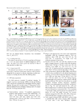
TaggedAPTARAFigure
           TaggedAPTARAEndHeat exposure on brain activity and cognitive function                                 235


           Fig. 1. Experimental design and task-based fMRI protocols. (A) For EX trial, participants were given ambient temperature drink after their first MRI scan, followed by
           a treadmill run until they achieved 39.5˚C. The trial ended with a second MRI scan after the run. PA trial shares the same protocol as EX except participants were
           passively heated in a water bath at 41˚C until they achieved 39.5˚C. Exertional hyperthermia with CL shares the same protocol as EX except participantsingested
           blended ice instead of ambient temperature (25˚C) drinks. CT excludes any hyperthermia-inducing interventions (exercise/immersion) where participants rested in a
           sitting position and ingested ambient temperature drinks. (B) Insulative thermal inner wear to limit heat loss and absorb sweat. (C) Water-perfused thermosuit with
           circulating water at 41˚C to restrict heat loss in scanner. (D) External water pump with insulated tubes. (E) Step ergometer (denoted by red box) for the conduct of plan-
           tarflexion task in MRI scanner. (F) Motor task-based fMRI scan where participants were asked to perform 10 sets of plantarflexion with rest intervals of 1 min, repeated
           for 12 blocks. (G) Cognitive task-based fMRI scan where participants performed the Stroop color-naming task for 1 min followed by 1-min fixation task, repeated for 4
           blocks. CL = cooling trial; CT = control trial; EX = exertional hyperthermia trial; fMRI = functional magnetic resonance imaging; MRI = magnetic resonance imaging;
           MVC = maximal voluntary contraction; PA = passive hyperthermia trial; VO 2peak = peak oxygen consumption.TaggedAPTARAEnd

           41˚C by an external heater. Immersion was terminated   TaggedAPTARAEndTaggedAPTARAP(b)  Single voxel spectroscopy with weak water suppression;
                                                                     bandwidth = 2000 Hz, acquisition duration = 1024 ms,
           when T re hit 39.5˚C.TaggedAPTARAEnd
                                                                     repetition time = 1500 ms, echo time = 30 ms, flip
                                                                     angle = 90˚, 32 acquisitions. The single voxel was
           TaggedAPTARAP2.2.3. Cooling trial (CL)TaggedAPTARAEnd
             TaggedAPTARAPThe protocol was similar to EX trial except that well-blended  centered at primary motor cortex.TaggedAPTARAEnd
           ice (1˚C) was given instead of ambient temperature (25˚C)  TaggedAPTARAP(c) Pseudo-continuous ASL with multi-slice single-shot echo
           water. The mass of ice slurry given was identical to that of  planar imaging sequence; 21 slices with 4 mm thickness,
                                                                     field of view = 256 mm £ 256 mm, matrix size = 64 £ 64,
           water provided in other trials.TaggedAPTARAEnd
                                                                     repetition time = 4000 ms, echo time = 9.2 ms, flip
                                                                     angle = 90˚, 20 label/control pairs, scanning time = 6 min,
           TaggedAPTARAP2.2.4. Control trial (CT)TaggedAPTARAEnd
             TaggedAPTARAPThe protocol was similar to PA trial with the exception of  label offset = 124 mm and post-label delay = 1500 ms.TaggedAPTARAEnd
           warm water immersion. Participants rested in a sitting position  TaggedAPTARAP(d) Task (motor)-based functional MRI (fMRI) with BOLD
           during the 60-min period (or shorter, depending on prior exer-  contrast using a gradient-echo echo planar imaging pulse
                                                                     sequence; 36 slices with 4-mm slice thickness, repetition
           cise/passive heating duration) after the drinking phase.TaggedAPTARAEnd
                                                                     time = 2000 ms, echo time = 30 ms, field of view = 256
                                                                     mm £ 256 mm, matrix size = 64 £ 64. During imaging,
           TaggedAPTARAH22.3. MRI data acquisitionTaggedAPTARAEnd
                                                                     the participants were placed in a supine position with the
                                                           1
             TaggedAPTARAPThe MRI protocols included T1-weighted imaging,  H
                                                                     head immobilized. The right foot was placed on a pedal
           magnetic resonance spectroscopy (MRS), arterial spin labeling
                                                                     ergometer (Trispect module; Ergospect, Innsbruck,
           (ASL), and blood oxygenation level-dependent (BOLD) contrast
                                                                     Austria), where participants performed dorsi- and plantar-
           imaging. All scans were recorded using a 3T Magnetom TrioTim
                                                                     flexion (Fig. 1E). Each task lasted for 20 s, with each
           (Siemens Medical Systems, Erlangen, Germany) and a 32-channel
                                                                     flexion performed at 2-s intervals with verbal instructions,
           head coil. The image protocols were:
                                                                     and was followed by a rest period of 40 s. This constitutes
                                                                     1 task block (1 min), and the process was repeated for 12
            TaggedAPTARAEndTaggedAPTARAP(a) High-resolution isotropic T1-weighted magnetization  blocks (12 min). The pedal resistance was pseudo-
               prepared rapid gradient recalled echo (MPRAGE); 176   randomly assigned at 20%, 40%, 60%, 80% of their pre-
               slices, 1 mm thickness, sagittal acquisition, field of
                                                                     determined maximal voluntary contraction (Fig. 1F).TaggedAPTARAEnd
               view = 256 mm £ 256 mm, matrix size = 256 £ 256,   TaggedAPTARAP(e) Task (cognitive)-based fMRI with BOLD contrast using
               repetition time = 1950 ms, echo time = 3.06 ms, inversion  a gradient-echo echo planar imaging pulse sequence;
               time = 900 ms, flip angle = 9˚.TaggedAPTARAEnd         36 slices with 4 mm slice thickness, repetition