
Page 119 - 《运动与健康科学》(英文)2024年第2期
P. 119
TaggedAPTARAFigure
TaggedAPTARAEndHeat exposure on brain activity and cognitive function 241
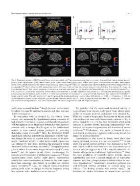
Fig. 6. Functional activation (BOLD) during Stroop color-naming task. (A) Task-related activation (task vs. no-task). Activated brain clusters include superior-
parietal gyrus, supramarginal gyrus, superior-frontal gyrus, caudal middle frontal gyrus, rostral middle frontal gyrus, lateral orbitofrontal cortex, medial orbito-
frontal cortex, lateral occipital cortex, superior temporal sulcus, isthmus cingulate cortex, and pars opercularis. (B) Normalized percent BOLD change (relative to
time-matched CT values) in baseline (S1) and post-intervention (S2) scans in left and right hemispheres: superior-temporal clusters. Bars represent the mean, and
lines represent the SD. Each symbol represents a value from an individual participant (n = 11). Significant difference between trials is denoted by asterisk (* p <
0.05). (C) PA vs. CT post scans comparison for task-related activation (task vs. no-task). Significant brain clusters (p < 0.05) include precentral gyrus, superior
parietal gyrus, superior temporal gyrus. (D) PA vs. CT post scans comparison for incongruent vs. congruent stimuli. Significant brain cluster in left hemisphere
lateral occipital cortex. The color scale in A, C, and D represents the cluster significance (-log(p)) where 4.00 denotes p < 0.0001, 1.33 denotes p < 0.05; red
gradient denotes increased activity while blue gradient denotes decreased activity. BOLD = blood oxygenation level-dependent; CL = cooling trial; CT = control
trial; EX = exertional hyperthermia trial; LH = left hemisphere; PA = passive hyperthermia trial; RH = right hemisphere.TaggedAPTARAEnd
39
and to preserve neural function, though the exact mechanism(s) TaggedAPTARAPWe postulate that the suppressed functional activity in
by which this could be mediated is unclear and, thus, warrants motor-related brain regions could result from altered excit-
ability or transient neuronal dysfunction with elevated T br .
further investigation.TaggedAPTARAEnd
TaggedAPTARAPIn conjunction with an elevated T br , the cortical motor While the effects of temperature fluctuations on human neural
activity was suppressed by hyperthermia during execution of function have not been well-characterized, variation of T br in
high-intensity motor tasks. However, a similar deficit was absent animal models by 1˚C3˚C has been reported to affect neural
with the ingestion of ice. High force exertion has been shown to function at multiple levels, including transmembrane ionic
associate with a stronger BOLD signal in the motor-related transport, action potential generation, and properties of passive
cortices as more cortical neurons participate in generating membrane. 46 Furthermore, heat stroke is known to cause
descending motor commands. 40 Here, the diminished BOLD neurological dysfunction in humans, underscoring the adverse
47
signal likely indicates a downstream impairment to motor func- impact on neuronal function. TaggedAPTARAEnd
tion, though the extent of physical deficit remains to be deter- TaggedAPTARAPHyperthermia is also known to impair brain blood flow
mined. Functional activity in other motor-related regions was during prolonged exercise in a hot environment. 48,49 In our
also affected by heat stress, including the paracentral lobule study, the global cerebral perfusion was depressed in the PA
41
implicated for lower extremity movement, the superior-parietal trial, which could be associated with the vasoconstriction of
gyrus involved in visuomotor integration and control of move- cerebral arteries triggered by a fall in arterial carbon dioxide
ments, 42 and the superior-frontal cluster encompassing the tension with hyperthermia-induced hyperventilation. 50 Given
43
premotor area for execution of skilled action sequences. Lower the lower production of metabolic carbon dioxide, passive
superior-frontal cortex activity under passive hyperthermia hyperthermia can elicit a greater magnitude of hypocapnic
could affect motor coordination as lesions in the same region response and, thus, a larger perfusion deficit relative to exer-
have been associated with deficits in complex and bilateral tional hyperthermia. 51 Interestingly, subcortical perfusion was
motor movements. 44 Last, the reduced lateral-orbitofrontal well-preserved whereas perfusion in cortical areas was
activity could relate to changes in affective responses during reduced. This finding concurs with another study 16 that
45
heat exposure, which modulate exercise tolerance. TaggedAPTARAEnd demonstrated similar reductions in sensorimotor perfusion