
Page 21 - 《水产学报》2025年第10期
P. 21
洪佳乐,等 水产学报, 2025, 49(10): 109602
400 control 0.5 mg/kg 1 mg/kg 1.5
a 2 mg/kg 4 mg/kg
b b c 1.0 a
300
量化长度/µm quantized length 200 d b b a 绒毛相对长度/µm villi relative length 0.5 b c
c
100
d
d c c b a dcd c b a
0 0
LMF WMF WLP WSM
肠道的形态结构 control 1 mg/kg 4 mg/kg
structural morphology of the intestine
不同处理组
different treatment groups
(a) (b)
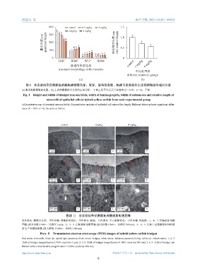
图 1 各实验组杂交黄颡鱼后肠黏膜褶皱高度、宽度,固有层宽度、黏膜下层宽度和上皮细胞微绒毛相对长度
(a) 肠道黏膜褶皱量化图;(b) 上皮细胞微绒毛长度的定量分析; 不同上标字母表示差异显著 (P < 0.05,n = 6),下同
Fig. 1 Height and width of hindgut mucosal folds, width of lamina propria, width of submucosa and relative length of
microvilli of epithelial cells in hybrid yellow catfish from each experimental group
(a)Quantitative map of intestinal mucosal folds; (b)quantitative analysis of epithelial cell microvillus length; Different letters indicate significant differ-
ences (P < 0.05, n = 6), the same as bellow.
control 1 mg/kg 4 mg/kg
1 μm 1 1 μm 4 1 μm 7
500 nm 2 500 nm 5 500 nm 8
200 nm 3 200 nm 6 200 nm 9
图版 Ⅱ 各实验组杂交黄颡鱼后肠透射电镜图像
红色箭头. 微绒毛长度;黑色尖端. 顶端紧密连接;黑色箭头. 桥粒;白色箭头. 下方黏附连接;白色尖端. 线粒体;1,4,7. 后肠透射电镜
图像 (放大倍数 2 900×;比例尺 1 μm);2,5,8. 后肠透射电镜图像 (放大倍数 9 300×;比例尺 500 nm);3,6,9. 后肠上皮细胞线粒体的透
射电子显微镜图像 (放大倍数 13 000×;比例尺 200 nm)
Plate Ⅱ Transmission electron microscopy (TEM) images of hybrid yellow catfish hindgut
Red arrow. microvilli; black tip. apical tight junctions; black arrow. bridges; white arrow. adherens junctions below; white tip. mitochondria; 1, 4, 7.
TEM of hindgut (magnification 2 900×; scale bar 1 μm); 2, 5, 8. TEM of hindgut (magnification 9 300×; scale bar 500 nm); 3, 6, 9. TEM of hindgut epi-
thelium cells’s mitochondria (magnification 13 000×; scale bar 200 nm).
https://www.china-fishery.cn 中国水产学会主办 sponsored by China Society of Fisheries
6