Page 23 - 《水产学报》2025年第8期
P. 23

黄泽翔,等                                                                 水产学报, 2025, 49(8): 089102

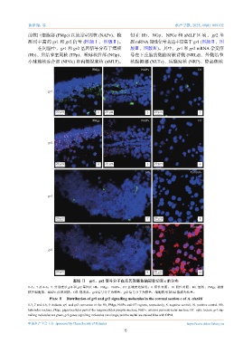
              前核巨细胞部 (PMgc) 以及前室周核 (NAPv),检                    但 在  Hb、 NGp、 NPGc 和     nMLF  区 域 , gr2  基
              测到丰富的       gr1  和  gr2  信号 (图版Ⅰ,图版Ⅱ)。           因  mRNA  阳性信号表达丰度高于          gr1 (图版Ⅱ,图
                   在间脑中,gr1      和  gr2  基因信号分布于缰核             版Ⅲ,图版Ⅳ)。其中,gr1           和  gr2 mRNA  杂交信
              (Hb)、后结节室周核 (TPp)、嗅球核后部 (NGp)、                   号在下丘脑的侧隐窝核背侧 (NRLd)、外侧结节
              小球前核连合部 (NPGc) 和内侧纵束核 (nMLF)。                    核腹侧部 (NLTv)、后隐窝核 (NRP)、隆起侧核


                                             PMgc                   NAPv                      OT


                        gr1


                           20 μm               1   20 μm              2   20 μm                3

                                             PMgc                   NAPv                      OT


                        gr2


                           20 μm               4   20 μm              5   20 μm                6

                                                                      Hb                   Control−


                        gr1


                           20 μm                                      7   20 μm                8

                                                                      Hb                   Control+


                        gr2


                           20 μm                                      9   20 μm                10

                                    图版 Ⅱ    gr1、gr2  信号分子在克氏双锯鱼脑冠状切面           c 的分布
              1~3,7  及  4~6,9. 分别表示  gr1  和  gr2  基因在  Hb、PMgc、NAPv、OT  区域表达情况,8. 阴性对照,10. 阳性对照。Hb. 缰核,PMgc. 视前
              核巨细胞部,NAPv. 前室周核,OT. 视顶盖。gr1     信号分子为绿色,gr2    信号分子为橙色,细胞核用       DPAI 染成为蓝色。
                        Plate Ⅱ Distribution of gr1 and gr2 signalling molecules in the coronal section c of A. clarkii
              1-3, 7 and 4-6, 9. indicate gr1 and gr2 expression in the Hb, PMgc, NAPv and OT regions, respectively, 8. negative control, 10. positive control. Hb.
              habenular nucleus, PMgc. gigantocellular part of the magnocellular preoptic nucleus, NAPv. anterior periventricular nucleus, OT. optic tectum. gr1 sig-
              nalling molecules are green, gr2 genes signalling molecules are orange, and the nuclei are stained blue with DPAI.

              中国水产学会主办  sponsored by China Society of Fisheries                          https://www.china-fishery.cn
                                                            5
   18   19   20   21   22   23   24   25   26   27   28