Page 89 - 《摩擦学学报》2021年第3期
P. 89

378                                     摩   擦   学   学   报                                 第 41 卷

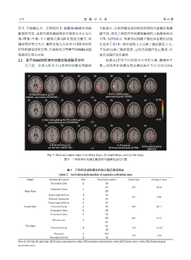
            信号. 在接触压力一定的情况下,根据图6触摸时的接                          大脑脑区,分别对触觉感知纹理形状的大脑激活数据
            触面积可知,皮肤内部机械感受体所受应力大小为尖                            做平均,得到三种纹理形状激发触感的大脑横状面切
            角>圆角>平角,其中触摸尖角试样时的应力最大. 机                          片图,如图9所示. 每种形状刺激下激活体素群的详细
            械感受体受力大小,最终表现为人体对不同纹理形状                            信息列于表2中,表中团块大小反映了激活脑区大小,

            特征的触觉感知差异,后面将结合摩擦学和fMRI试验                          平均t值反映了激活强度,正值代表脑区的正激活,负
            数据进行深入讨论.                                          值代表脑区的负激活.

            2.2    基于fMRI的纹理形状激发触感脑区分析                             根据表2中每个团块的大小可得尖角、圆角和平
                为了进一步深入研究不同纹理形状激发的触感                           角三种纹理形状激发的总激活脑区大小分别为364、


                  25         30        35        40         45        50        55         60


                   R.Supramarginal gyrus/                            L.Prccentral gyrus/
                 rolandic operculum (SI/SII/MI)           (a)      postcentral gyrus (SI/SII)
                  25         30        35        40         45        50        55         60     12.98


                           L.Supramarginal gyrus/Precentral gyrus/                                −8.92
                               postcentral gyrus (SI/SII/MI)  (b)
                  25         30        35        40         45        50        55         60


                                L.Postcentral gyrus/                      L.Precuneus
                               parital lobe (SI/SII/MI)   (c)        R.Paracentral lobuls (SI/MI)  R.Precentral gyrus
                                                                                             (SI/SSA)

                               Fig. 9  Brain activation maps of (a) sharp shape,(b) round shape,and (c) flat shape
                                         图 9    三种纹理形状激发触感的大脑横状面切片图


                                           表 2    三种形状试样激发的独立脑区激活信息
                                      Table 2    Activation information of separate activation area

                  Sample        Anatomical location  Side     Functional location  Cluster size  Average t-value
                                 Precentral Gyrus    L             SI
                                                                   MI              263            10.34
                                Postcentral Gyrus    L
                Sharp shape                                        SII
                               Supramarginal Gyrus   R             SI
                                                                                   101             8.86
                                Rolandic Operculum   R             SII
                               Supramarginal Gyrus   L             SI
                Round shape      Precentral Gyrus    L             MI              268            10.11
                                Postcentral Gyrus    L             SII
                                Postcentral Gyrus    L             SI
                                                                   MI              266            11.33
                                  Parietal Lobe      L
                                                                   SII
                 Flat shape                                        MI
                                 Precentral Gyrus    R                             104            -14.62
                                                                   SI
                                   Precuneus         L            SSA
                                                                                   109             -7.46
                                Paracentral Lobule   R             SI
            Note:L-left brain;R-right brain;SI-Primary somatosensory cortex;SII-Secondary somatosensory cortex;MI-Primary motor cortex;SSA-Somatosensory
            association cortex.
   84   85   86   87   88   89   90   91   92   93   94