
Page 130 - 《运动与健康科学》(英文)2024年第2期
P. 130
TaggedAPTARAFigure TaggedAPTARAEnd252 C.S. Norevik et al.
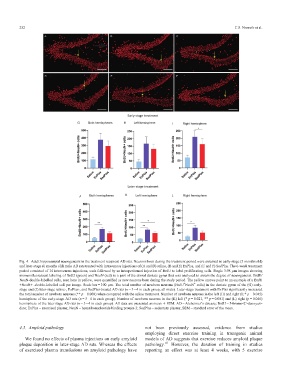
Fig. 4. Adult hippocampal neurogenesis in the treatment recipient AD rats. Neurons born during the treatment period were assessed in early-stage (3 months old)
and later-stage (6 months old) male AD rats treated with intravenous injections of (A and D) saline, (B and E) ExPlas, and (C and F) SedPlas. The 6-week treatment
period consisted of 14 intravenous injections, each followed by an intraperitoneal injection of BrdU to label proliferating cells. Single 3.98 mm images showing
immunofluorescent labeling of BrdU (green) and NeuN (red) in a part of the dorsal dentate gyrus that was analyzed to assess the degree of neurogenesis. BrdU/
NeuN-double-labelled cells, seen here in yellow, were quantified as new neurons born during the study period. The yellow arrows point to an example of a BrdU
+
+
+NeuN+ -double-labelled cell per image. Scale bar = 100 mm. The total number of newborn neurons (BrdU NeuN cells) in the dentate gyrus of the (G) early-
stage and (J) later-stage saline-, ExPlas-, and SedPlas-treated AD rats (n =34 in each group, all male). Later-stage treatment with ExPlas significantly increased
the total number of newborn neurons (** p = 0.008) when compared with the saline treatment. Number of newborn neurons in the left (H) and right (I; * p = 0.045)
hemisphere of the early-stage AD rats (n =34 in each group). Number of newborn neurons in the (K) left (* p = 0.021, ** p = 0.001) and (L) right (p = 0.046)
hemisphere of the later stage AD rats (n =34 in each group). All data are presented as mean § SEM. AD = Alzheimer’s disease; BrdU = 5-bromo-2’-deoxyuri-
dine; ExPlas = exercised plasma; NeuN = hexaribonucleotide binding protein-3; SedPlas = sedentary plasma; SEM = standard error of the mean.TaggedAPTARAEnd
not been previously assessed, evidence from studies
TaggedAPTARAH24.3. Amyloid pathologyTaggedAPTARAEnd
employing direct exercise training in transgenic animal
TaggedAPTARAPWe found no effects of plasma injections on early amyloid models of AD suggests that exercise reduces amyloid plaque
plaque deposition in later-stage AD rats. Whereas the effects pathology. 41 However, the duration of training in studies
of exercised plasma transfusions on amyloid pathology have reporting an effect was at least 4 weeks, with 5 exercise