
Page 32 - 《渔业研究》2026年第3期
P. 32
第 3 期 戴景辉等: 患病杂交鲟鲟疱疹病毒 3 型与海豚链球菌的共检出、病理特征及药敏分析 325
a) 鳃 Gill b) 大脑 Brain
A A’ B B’
c) 脊椎旁肌 Paraspinal muscles d) 腹腔 Abdominal cavity
★
C C’ D D’
e) 脾脏 Spleen f) 肠道 Intestine
E E’ F F’
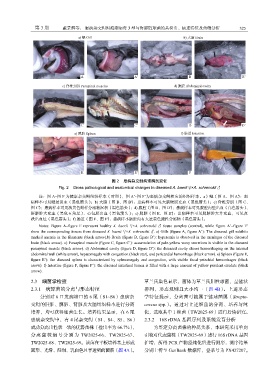
图 2 患病杂交鲟病理解剖变化
Fig. 2 Gross pathological and anatomical changes in diseased A. baerii ♀×A. schrenckii ♂
注:图 A~图 F 为健康杂交鲟组织样本(对照) ,图 A’~图 F’为患病杂交鲟相应的组织样本。a)鳃(图 A、图 A’) :患
病样本可见鳃丝贫血(黑色箭头) ;b) 大脑(图 B、图 B’) :患病样本可见大脑硬膜充血(黑色箭头) ;c) 脊椎旁肌(图 C、
图 C’) :患病样本可见淡黄色蜡样分泌物沉积(黑色箭头) ;d) 腹腔(图 D、图 D’) :患病样本可见腹腔内壁出血(白色箭头) 、
肝脏肿大充血(黑色五角星) 、心包膜出血(黑色箭头) ;e) 脾脏(图 E、图 E’) :患病样本可见脾脏肿大并充血,可见点
状出血灶(黑色箭头) ;f) 肠道(图 F、图 F’) :患病样本肠腔内有大量黄色脓性分泌物(黑色箭头) 。
Notes: Figure A–figure F represent healthy A. baerii ♀×A. schrenckii ♂ tissue samples (control), while figure A’–figure F’
show the corresponding tissues from diseased A. baerii ♀×A. schrenckii ♂. a) Gills (figure A, figure A’): The diseased gill exhibits
marked anemia in the filaments (black arrow).b) Brain (figure B, figure B’): hyperemia is observed in the meninges of the diseased
brain (black arrow). c) Paraspinal muscle (figure C, figure C’): accumulation of pale-yellow waxy secretions is visible in the diseased
paraspinal muscle (black arrow). d) Abdominal cavity (figure D, figure D’): the diseased cavity shows hemorrhaging on the internal
abdominal wall (white arrow), hepatomegaly with congestion (black star), and pericardial hemorrhage (black arrow). e) Spleen (figure E,
figure E’): the diseased spleen is characterized by splenomegaly and congestion, with visible focal petechial hemorrhages (black
arrow). f) Intestine (figure F, figure F’): the diseased intestinal lumen is filled with a large amount of yellow purulent exudate (black
arrow).
2.3 细菌学检查 革兰氏染色显示,菌体为革兰氏阳性球菌,呈链状
2.3.1 病原菌的分离与形态特征 排列,形态规则且大小均一(图 4B) 。上述形态
分别对 6 口发病塘口的 6 尾(S1~S6)患病杂 学特征提示,分离菌可能属于链球菌属(Strepto-
交鲟的肝脏、脾脏、肾脏及大脑组织样本进行分离 coccus spp.) 。通过对上述细菌的分离、培养与纯
培养,均可获得细菌生长。培养结果显示,在 6 尾 化,选取其中 1 株菌(TW2025-69)进行后续研究。
患病杂交鲟中,有 4 尾杂交鲟(S1、S4、S5、S6) 2.3.2 16S rDNA 基因序列及系统发育分析
成功分离出性状一致的优势菌株(检出率为 66.7%) , 为鉴定分离菌株的种属关系,本研究采用单向
分 离 菌 株 编 号 分 别 为 TW2025-66、 TW2025-67、 引物对代表菌株(TW2025-69)进行 16S rDNA 基因
TW2025-68、TW2025-69。该菌在平板培养基上形成 扩增,所得 PCR 产物经纯化后进行测序,测序结果
圆形、光滑、湿润、乳白色且半透明的菌落(图 4A)。 分别上传至 GenBank 数据库,登录号为 PX427207。