
Page 154 - 《水产学报》2026年第2期
P. 154
2 期 PASSMORE Roland Madziva,等:银灰半棱鳀仔稚鱼的脊柱和附肢骨骼发育 50 卷
Bp
pb
Ra
Fr
0.50 mm 1 0.50 mm 2 0.50 mm 3
ps
ps
Ra
Bp
ps
0.50 mm 4 1.00 mm 5 1.00 mm 6
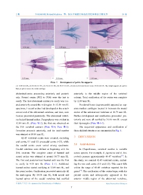
Plate Ⅴ Development of pelvic fin supports
(1) 12.89 mm SL; (2) 15.06 mm NL; (3) 18.22 mm SL; (4) 31.07 mm SL; (5)-(6) ventral and lateral views: 34.56 mm SL. Bp. basipterygium; pb. pelvic
bud; ps. pelvic scute; Ra. radial cartilage.
abdominal centra proceeding anteriorly and posteri- anteriorly to the middle region of the vertebral
orly. Preural centra (PU2 to PU4) were the last to column. Total ossification of the centra was complete
ossify. The first abdominal centrum to ossify was loc- by 12.89 mm SL.
ated anteriorly around the mid-region. In 12.89 mm SL Pre-dorsal bones (supraneurals) appeared as sep-
specimen, 3 neural arches had developed at the anteri- arate median cartilages located in between the neural
ormost end of the abdominal vertebrae, and later, ossi- arches of the anteriormost vertebrae at 14.73 mm SL.
fication proceeded posteriorly. The abdominal verteb- Further development and ossification proceeded pos-
rae lacked haemal arches. Parapophysis was evident in teriorly and were all ossified by 34.46 mm SL except
12.89 mm SL (Plate Ⅵ-2); the first one observed on their tip margins (Plate Ⅶ-1-3).
the 25th vertebral centrum (Plate Ⅵ-4, Plate Ⅶ-2): The sequential appearance and ossification of
formation proceeds anteriorly, and the total number these skeletal structures are summarized in Fig. 1.
was attained at 18.08 mm SL.
3 DISCUSSION
42-43 vertebral centra were counted, excluding
ural centra, U1 and U2: precaudal centra (=25), while
3.1 Axial skeleton
the caudal centra count varied among specimens.
Caudal vertebrae were defined as beginning with the In Clupeiformes, vertebral number is variable
26th centrum. The complete count of haemal and across species. For example, E. japonicus and E. het-
[22]
neural arches was attained at around 14.73 mm SL. erobola possess approximately 44-47 vertebrae . In
The first and posteriormost haemal arch was the first this study, we counted 42-43 vertebral centra, exclud-
to ossify in 9.58 mm SL (Plate Ⅰ-1). Additional ing the two ural centra (U1 and U2). This count falls
haemal arches started ossifying at 12.89 mm SL, and within the range of 41-43 vertebrae reported for the
[22]
the neural arches. Ossification proceeded anteriorly till genus . The ossification of the centra began with the
the mid-region. By 18.08 mm SL, both neural and preural centra and subsequently appeared in the
haemal spines of the caudal vertebrae had ossified anterior middle region of the abdominal vertebrae,
中国水产学会主办 sponsored by China Society of Fisheries https://www.china-fishery.cn
7