
Page 152 - 《水产学报》2026年第2期
P. 152
2 期 PASSMORE Roland Madziva,等:银灰半棱鳀仔稚鱼的脊柱和附肢骨骼发育 50 卷
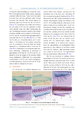
3.78 mm NL, then procartilage at 5.20 mm NL (Plate ossified dermal bone structure extending from the
Ⅲ-1). Subsequent proximal radials emerge, appearing anterolateral side of the body, and a thin dermal fin
embedded within the tissue. Central proximal radials fold developed and gradually thickened (Plate Ⅳ-1).
developed first, and then additional radials formed Subsequently, the radial fin plate developed, and rudi-
posteriorly and anteriorly. They become apparent in mentary coracoscapular cartilage was evident in 5.25
specimens larger than 6.08 mm NL. By 5.79 mm NL mm NL. This cartilage featured a small groove at the
(Plate Ⅲ-2), cartilaginous distal radials had formed at anterior margin and an extended, short, curved pos-
the mid region of the anal fin, appearing as small terior process. Around 6.08 mm NL, sequential seg-
round elements along with their corresponding fin mentation of the radial fin plate became noticeable. At
rays. Development proceeds in anterior and posterior
6.81 mm NL, a median crevice was evident and later
directions, and full count is reached by 10.20 mm SL.
followed by the emergence of two other crevices by
At 11.91 mm SL, the posteriormost proximal radial is
12.89 mm SL (Plate Ⅳ-2): two originate from the
associated with two fin rays (Plate Ⅲ-4-6). At 18.08
middle of the fin plate, and one median crevice
mm SL, there are 14 proximal radials, and 14 distal
extends from the anterior-ventral margin. These
radials. A developing stay bone appears originating
crevices developed in an inner-to-outer sequence.
from the posteriormost proximal radial and remain
Later, the supracleithrum and posttemporal formed
separated by a cartilaginous band at 34.56 mm SL
antero-dorsal to the cleithrum by ossification around
(Plate Ⅲ-6). Ossification of the proximal radials pro-
10.24 mm SL. However, their association became
ceeds posteriorly, commencing in specimens at
more evident at 12.89 mm SL. The supracleithrum
approximately 18.08 mm SL. At 30.42 mm SL, the
then articulated with the cleithrum (Plate Ⅳ-2-4).
innermost dorsal tip margins of the proximal radials
The fin rays developed as cartilage between
remain cartilaginous. Additionally, the corresponding
approximately 15.03 and 16.33 mm SL and were con-
ventral margins of the fin rays remain cartilaginous,
sistently observed in specimens from 15.03 to 34.46
with full segmentation evident at 34.56 mm NL. Distal
mm SL. These were the last fin rays to form. All pec-
radials remain as cartilaginous structures.
toral fin rays had ossified by 34.56 mm SL, with a
2.4 Pectoral fin
total count of 14 rays. The propterygium was posi-
At 3.78 mm NL, the cleithrum appeared as an tioned on the postero-dorsal part of the scapula and
Pr
Dr
Afa Pr
Dr
0.25 mm 1 0.25 mm 2 0.25 mm 3
Fr Sb
Pr Dr Sb
Fr
0.25 mm 1 mm 1 mm
4 5 6
Plate Ⅲ Development of the anal fin supports and fin rays
(1) 5.20 mm NL; (2) 5.79 mm NL; (3) 10.24 mm SL; (4) 11.91 mm SL; (5) 18.08 mm SL; (6) 34.56 mm SL. Afa. anal fin anlagen.
中国水产学会主办 sponsored by China Society of Fisheries https://www.china-fishery.cn
5