
Page 185 - 《水产学报》2025年第12期
P. 185
张金叶,等 水产学报, 2025, 49(12): 129414
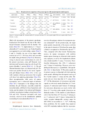
Tab. 1 Morphometrical comparison of the present species with morphologically similar species
PF
species name SL/μm SW/μm ST/μm PCL/μm PCW/μm sites of infection hosts locality references
turns
T. xiushanensis n. sp. 19.2 ± 1.1 10.0 ± 0.7 9.7 ± 0.5 8.0 ± 0.6 6.6 ± 0.3 4 - 5 gill filaments C. auratus China this study
(17.1 - 21.0) (8.7 - 12.1) (9.1 - 10.8) (6.9 - 9.1) (6.1 - 7.1)
T. wangi 20.2 9.9 9.3 10.1 6.5 6 - 7 gill filaments C. gibelio China [19]
(16.5 - 22.3) (9.1–10.8) (8.2 - 10.4) (8.4 - 11.2) (6.1 - 7.0)
T. hovorkai 20.4 9.8 8.5 10.8 8.9 6 - 7 gills, gallbladder, C. auratus, China [20]
(18.0 - 22.4) (7.2 - 12.0) (7.0 - 10.0) (7.2 - 14.4) (7.2 - 10.8) kidney C. carpio
T. wuhanensis 23.6 13.8 11.7 11.5 9.1 8 - 10 skin C. gibelio China [20]
(21 - 25) (12.0–15.5) (10.8 - 14.1) (9.6 - 12.7) (8.1 - 10.3)
T. hokiangensis 23.9 10.74 10.74 13.39 9.18 6 - 7 ureter, intestine C. carpio China [20]
(22.1 - 25.5) (9.35- 11.05) (9.35 - 11.9) (11.9 - 15.3) (8.5 - 9.35)
T. kitauei 33.4 15.0 - 16.8 7.4 8 - 10 intestine C. carpio Japan [21]
(31 - 35) (12 - 17) (14 - 18) (6 - 9)
Notes: SL. myxospore length; SW. myxospore width; ST. myxospore thickness; PCL. polar capsule length; PCW. polar capsule width; PF. polar
filament; -. no data.
filled with myxospores of the present specimens, served as the primary criterion for myxsporean spe-
localized and developed near the tips of the infec- cies delineation . In the present study, the single
[22]
ted gill filaments compared with the healthy fila- polar capsule characteristic of the species conforms
ments (Plate II-1 - 7). Approximately 2 - 5 myxo- to the typical character of Thelohanellus genus, thus
plasmodia of T. xiushanensis n. sp. flocked together [4, 20]
it firstly is recognized as Thelohanellus species .
and separated by cyst walls within a nidus (Plate II-
Then, compared with other known Thelohanellus
4 and 5). Notably, the size of the largest nidus
species, its mature myxospores with pyriform
reached up to 93.3 × 38.4 μm, which had exceeded
mature spores featuring an ellipsoidal polar capsule
the width of the gill filament (Plate II-5). Moreover,
and a epispore, as well as the morphometric dimen-
owing to physical space encroachment by cysts of
sion, closely resemble T. wangi, T. hovorkai, Thelo-
the present specimens, some gill filaments were
hanellus hokiangensis, Ma, 1998, T. wuhanensis
deformed and fractured comparing with the gill fila-
and T. kitauei in morphology. However, some mor-
ments of healthy C. auratus (Plate II-1 - 4). The gill
phological discrepancy is quantifiably distinguish-
filament basement layer and diseased old gill fila-
able (Tab. 1). Specifically: ① The polar capsule of
ments were pushed forward by the newly regener-
the present species is ellipsoidal with short-opening
ated gill filament tissue (Plate II-4). A few cyst
polar capsule, differing from the tapered opening of
walls ruptured, releasing myxospores that mingled
with those from adjoining plasmodium (Plate II-8). the of polar capsule T. wangi, and the polar fila-
Most myxoplasmodia, with nearly all mature ment coils are fewer than that of T. wangi (Plate
[19]
myxospores, were in advanced stages of infection I) . ② The dimensions of myxospores and polar
(Plate II-7). Consequent to the damage from the capsule are obviously smaller than those of T. hoki-
infection, the gill filaments, especially around the angensis, T. wuhanensis and T. kitauei. ③ Although
myxoplasmodia, exhibited obvious hyperplasia and the myxospore dimensions are nearly similar with
fusion, and the termini of the infected gill filaments those of T. hovorkai, polar capsule dimensions are
exhibited vacuolization leading to detachment (Plate distinctly smaller. ④ The number of polar filament
II-4 and 9-10). Numerous eosinophil cells were coils is less than those of T. hovorkai, T. hokiangen-
obviously observed in the interlamellar epithelia sis, T. wuhanensis and T. kitauei. Independent the
surrounding the myxoplasmodia (Plate II-11). widely accepted intraspecific variance range for
[23]
SSU rDNA in myxobolids is ≤ 1% . Following
3 DISCUSSION
these guidelines, the two equally long sequences
Morphological characters have historically with 100% similarity indicates that the myxospor-
中国水产学会主办 sponsored by China Society of Fisheries https://www.china-fishery.cn
7