
Page 184 - 《水产学报》2025年第12期
P. 184
张金叶,等 水产学报, 2025, 49(12): 129414
S
F
S F
GA
F S
GR
1 mm 1 250 μm 2 50 μm 3
P P
P P
P P
P
* *
P P
P P
P P
P *
* P P
* P
* * P
P P
1 mm 4 100 μm 5 10 μm 6
10 μm 7 50 μm 8 100 μm 9
100 μm 10 20 μm 11
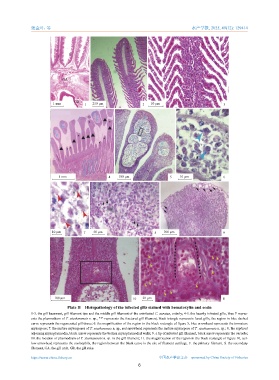
Plate II Histopathology of the infected gills stained with hematoxylin and eosin
1-3. the gill basement, gill filament tips and the middle gill filament of the uninfected C. auratus, orderly; 4-5. the heavily infected gills, blue P repres-
ents the plasmodium of T. xiushanensis n. sp., "*" represents the fractured gill filament, black triangle represents fused gills, the region in blue dashed
curve represents the regenerated gill tissue; 6. the magnification of the region in the black rectangle of figure 5, blue arrowhead represents the immature
myxospore; 7. the mature myxospores of T. xiushanensis n. sp., red arrowhead represents the mature myxospore of T. xiushanensis n. sp.; 8. the ruptured
adjoining myxoplasmodia, black arrow represents the broken myxoplasmodial walls; 9. a tip of infected gill filament, black arrow represents the vacuole;
10. the location of plasmodium of T. xiushanensis n. sp. in the gill filament; 11. the magnification of the region in the black rectangle of figure 10, yel-
low arrowhead represents the eosinophils, the region between the black curve is the site of filament cartilage. F. the primary filament, S. the secondary
filament, GA. the gill arch, GR. the gill rake.
https://www.china-fishery.cn 中国水产学会主办 sponsored by China Society of Fisheries
6