
Page 461 - 《软件学报》2024年第6期
P. 461
胡凯 等: 基于端到端深度神经网络和图搜索的 OCT 图像视网膜层边界分割方法 3037
2
(Hunan Provincial Key Laboratory of Intelligent Computing and Language Information Processing (Hunan Normal University), Changsha
410081, China)
3
(Key Laboratory of Medical Imaging and Artificial Intelligence of Hunan Province (Xiangnan University), Chenzhou 423000, China)
Abstract: The morphological changes in retina boundaries are important indicators of retinal diseases, and the subtle changes can be
captured by images obtained by optical coherence tomography (OCT). The retinal layer boundary segmentation based on OCT images can
assist in the clinical judgment of related diseases. In OCT images, due to the diverse morphological changes in retina boundaries, the key
boundary-related information, such as contexts and saliency boundaries, is crucial to the judgment and segmentation of layer boundaries.
However, existing segmentation methods lack the consideration of the above information, which results in incomplete and discontinuous
boundaries. To solve the above problems, this study proposes a coarse-to-fine method for the segmentation of retinal layer boundary
in OCT images based on the end-to-end deep neural networks and graph search (GS), which avoids the phenomenon of “faults” common
in non-end-to-end methods. In coarse segmentation, the attention global residual network (AGR-Net), an end-to-end deep neural network, is
proposed to extract the above key information in a more sufficient and effective way. Specifically, a global feature module (GFM) is
designed to capture the global context information of OCT images by scanning from four directions of the images. After that, the channel
attention module (CAM) and GFM are sequentially combined and embedded in the backbone network to realize saliency modeling of
context information of the retina and its boundaries. This effort effectively solves the problem of wrong segmentation caused by retina
deformation and insufficient information extraction in OCT images. In fine segmentation, a GS algorithm is adopted to remove isolated
areas or holes from the coarse segmentation results obtained by AGR-Net. In this way, the boundary keeps a fixed topology, and it is
continuous and smooth, which further optimizes the overall segmentation results and provides a more complete reference for medical
clinical diagnosis. Finally, the performance of the proposed method is evaluated from different perspectives on two public datasets, and the
method is compared with the latest methods. The comparative experiments show that the proposed method outperforms the existing
methods in terms of segmentation accuracy and stability.
Key words: optical coherence tomography (OCT) image; segmentation of retinal layer boundary; residual neural network; attention; graph
search (GS)
1 引 言
眼部疾病是人类常见疾病之一, 尤其是视网膜疾病, 对视功能损害巨大, 是影响人类视觉健康的重要因素. 随
着疾病类型和患者数量的增多, 临床诊断往往借助医学影像技术以更高的效率为患者提供诊察结果和治疗建议.
[1]
光学相干断层扫描 (optical coherence tomography, OCT) 是近年来发展迅速的新型层析成像技术, 填补了传统超
声和显微镜成像之间的空白 [2] , 且由于其对光具有高敏感度, 成像也具有较高的分辨率, 因此被广泛应用于眼部结
构成像及相关疾病的诊断及筛查等 [3−5] . 由于眼部疾病会导致视网膜层结构出现显著性变化, 临床医师往往通过观
察视网膜 OCT 图像中的层边界来帮助分析眼部疾病的病变程度, 通过视网膜层的厚度变化进行定量或定性分析
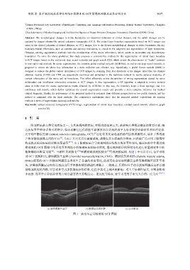
能够辅助诊断青光眼 [6] 、年龄性黄斑病变 [7,8] 和糖尿病视网膜病变 [9] 等视网膜疾病. 从图 1 中可以看出, 与正常眼
部 OCT 图像相比, 脉络膜新生血管 (choroidal neovascularization, CNV)、糖尿病黄斑水肿 (diabetic macular edema,
DME) 病变的 OCT 图像视网膜层边界发生了显著的形态变化, 此类变化会使视网膜层边界变得模糊不清甚至消
失, 使得视网膜层边界的分割成为医学图像领域的挑战性难题之一. 临床上, 医师往往手动分割视网膜层边界以辅
助眼部疾病的判断, 尽管这种方式能获得较好的结果, 但由于其受主观因素影响、对人工依赖性较大, 导致诊断效
率较低. 而基于计算机算法的自动分割方法具有精度高、速度快等特点, 近年来受到了研究人员的广泛关注 [10,11] .
(a) 脉络膜新生血管 (b) 糖尿病黄斑水肿 (c) 正常眼部
图 1 病变与正常眼部的 OCT 图像