
Page 41 - 《运动与健康科学》(英文)2024年第2期
P. 41
TaggedAPTARAEndEffect of exercise on locomotor system 163
TaggedAPTARAEnd by the upregulation of transient sodium channels 23,29,30 or the
31
downregulation of K(DR) channels. Furthermore, excitation of
motoneurons is mediated by the activation of PICs generated by
Chen and Dai (2022) 15 TaggedAPTARAEndTaggedAPTARAEnd Ge and Dai (2020) 16 TaggedAPTARAEndTaggedAPTARAEnd Chen and Dai (2022) 15 TaggedAPTARAEndTaggedAPTARAEnd Ge and Dai (2020) 16 TaggedAPTARAEndTaggedAPTARAEnd Beaumont and Gardiner (2002) 11 TaggedAPTARAEndTaggedAPTARAEnd Krutki et al. (2015) 45 TaggedAPTARAEndTaggedAPTARAEnd Woodrow et al. (2013) 92 TaggedAPTARAEndTaggedAPTARAEnd Li et al. (2013) 93 TaggedAPTARAEndTaggedAPTARAEndTaggedAPTARAEnd L-
3235
and amplifi-
for repetitive discharge of spinal motoneurons
36
cation of synaptic inputs from excitatory reflexes.
It is noted
ReferenceTaggedAPTARAEndTaggedAPTARAEndTaggedAPTARAEndTaggedAPTARATbody that Vth and PICs can be modulated by monoamines during
acute exercise. It has been shown previously that serotonergic
(5-HT) receptors (5-HT 1A ,5-HT 2A , and 5-HT 7 ) are co-expressed
in the spinal neurons, which are activated by electrical stimula-
tion of the mesencephalic locomotor region in cats, suggesting
Change in channel conductance, kinetics, or proteinTaggedAPTARAEnd that monoaminergic fibers contact neurons involved in generating
37
locomotion.
Furthermore, previous studies have reported that
38
and enhances PICs in the spinal
serotonin hyperpolarizes Vth
"TaggedAPTARAEnd
"TaggedAPTARAEnd
"TaggedAPTARAEnd
"TaggedAPTARAEnd
"TaggedAPTARAEnd
"TaggedAPTARAEnd
"TaggedAPTARAEnd
#TaggedAPTARAEnd
neurons of rodents. 39 These studies suggest that monoamines
such as serotonin can play an essential role in modulating
Ion channel modulatedTaggedAPTARAEnd Persistent sodium channelTaggedAPTARAEnd Persistent sodium channelTaggedAPTARAEnd L-type calcium channelTaggedAPTARAEnd L-type calcium channelTaggedAPTARAEnd neuronal excitability during acute exercise.TaggedAPTARAEnd
TaggedAPTARAPAlthough it is impossible to directly isolate motoneurons
from the human spinal cord for recording, researchers have
used non-invasive measurement techniques to study the
activity of motoneuron pools during different motor tasks
K(Ca)TaggedAPTARAEnd
K(Ca)TaggedAPTARAEnd
K(Ca)TaggedAPTARAEnd
K(Ca)TaggedAPTARAEnd
(acute exercise). In several studies of human motoneurons,
they have investigated the task-dependence of spinal moto-
neuron excitability during arm cycling by comparing the
responses evoked during the cycling to those evoked during
4042
position- and intensity-matched isometric contractions.
Abbreviations: 5-HT = serotonin; K(Ca) = calcium-activated potassium channel; VO 2max = maximal oxygen uptake.TaggedAPTARAEnd
motoneuron excitability is present but depends on the arm
14,40
Neuron affectedTaggedAPTARAEnd Lamina X interneuronTaggedAPTARAEnd Midbrain 5-HT neuronTaggedAPTARAEnd Lamina X interneuronTaggedAPTARAEnd Midbrain 5-HT neuronTaggedAPTARAEnd MotoneuronTaggedAPTARAEnd MotoneuronTaggedAPTARAEnd MotoneuronTaggedAPTARAEnd Cerebral artery smooth muscle cellTaggedAPTARAEnd Their findings suggest that the task-dependency in spinal
This task dependency in
position during arm cycling.
human spinal motoneurons during acute exercise has
some similarity to the state-dependent property in spinal
motoneurons during fictive locomotion in non-human
vertebrates.TaggedAPTARAEnd
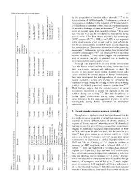
DurationTaggedAPTARAEnd 3 weeksTaggedAPTARAEnd 3 weeksTaggedAPTARAEnd 3 weeksTaggedAPTARAEnd 3 weeksTaggedAPTARAEnd 12 weeksTaggedAPTARAEnd 5 or 12 weeksTaggedAPTARAEnd 16 weeksTaggedAPTARAEnd 12 weeksTaggedAPTARAEnd TaggedAPTARAH13. Chronic exercise enhances neuronal excitabilityTaggedAPTARAEnd
TaggedAPTARAPThrough several studies in rats, it has been observed that the
electrophysiological properties of spinal motoneurons vary in
response to several different forms of chronic exercise. A
20 m/min, »50%55%VO 2max ,
regimen of “forced exercise” subjecting rats to 2 h of treadmill
60 min/day, 3 or 5 days/weekTaggedAPTARAEnd
27 m/min at a 10˚ incline for
1013 m/min, 60 min/day,
1013 m/min, 60 min/day,
1013 m/min, 60 min/day,
1013 m/min, 60 min/day,
training per day for up to 16 weeks caused their spinal moto-
neurons to experience significant hyperpolarization of RMP
and Vth, reductions in spike rise time, and decreases in FI
TaggedAPTARACaptionIon channel in response to exercise training in rodents. IntensityTaggedAPTARAEnd Exercise protocolTaggedAPTARAEnd TreadmillTaggedAPTARAEnd 6 days/weekTaggedAPTARAEnd TreadmillTaggedAPTARAEnd 6 days/weekTaggedAPTARAEnd TreadmillTaggedAPTARAEnd 6 days/weekTaggedAPTARAEnd TreadmillTaggedAPTARAEnd 6 days/weekTaggedAPTARAEnd SpontaneousTaggedAPTARAEnd Exercise wheelsTaggedAPTARAEnd Compensatory overloadTaggedAPTARAEnd Bilateral tenotomyTaggedAP
10
Rats given access to an exercise wheel for 1220
slope.
motoneurons to experience a similar hyperpolarization of
RMP and Vth and lowering of FI slopes in addition to
increases in AHP amplitude and a leftward shift of FI
11,43
Rats that underwent a “resistance-type exercise”
curves.
regimen for 1 h per day, 5 days per week for 5 weeks caused
their spinal motoneurons to experience a decrease in spike rise
44
Further-
time and rheobase and an increase in FI slope.
more, rats subjected to compensatory muscle overload through
enced an increase in AHP amplitude and Rin and a decrease in
Table 2 SpeciesTaggedAPTARAEnd MouseTaggedAPTARAEnd MouseTaggedAPTARAEnd MouseTaggedAPTARAEnd MouseTaggedAPTARAEnd RatTaggedAPTARAEnd RatTaggedAPTARAEnd RatTaggedAPTARAEnd RatTaggedAPTARAEnd tenotomy of synergistic muscles for up to 12 weeks experi-
45
Rats that underwent repeated
spike rise time and rheobase.
sessions of transcutaneous, trans-spinal direct current stimulation