
Page 209 - 《水产学报》2026年第01期
P. 209
1 期 管西涛,等:锦鲤寄生葡萄碘泡虫的形态特征、组织病理及分子系统发育 50 卷
3 μm 2
5 μm 1 3 μm 3
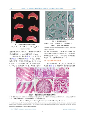
图版 Ⅰ 葡萄碘泡虫的孢子
10 mm
1. 孢囊内大量的孢子;2. 壳面观的孢子;3. 缝面观的孢子。
图 1 寄生锦鲤鳃的葡萄碘泡虫孢囊
Plate Ⅰ Spores of M. acinosus
Fig. 1 Plasmodia of M. acinosus infecting gills of
1. a number of spores from a plasmodium; 2. spore in frontal view;
C. carpio var. koi 3. spore in sutural view.
孢囊形状为圆形、椭圆形、长椭圆形或不规则形 0.2) μm, 4.4~5.6 μm], 大 极 囊 宽 [(2.8±0.1) μm,
状,孢囊长 0.4~1.0 mm,孢囊宽 0.3~0.6 mm。 2.5~3.0 μm];小极囊长 [(2.6±0.2) μm,2.3~3.0 μm],
小极囊宽 [(1.1±0.1) μm,1.0~1.3 μm]。大极囊内极
孢囊内存在大量孢子 (图版 Ⅰ-1),孢子壳面
丝为 6~7 圈,小极囊内极丝为 2~3 圈。
观 (图版 Ⅰ-2) 和缝面观 (图版 Ⅰ-3) 均呈长葡萄形,
孢质中存在 1 个明显的嗜碘泡。孢子长 [(11.3± 2.2 组织病理学特征描述
0.3) μm, 10.7~11.7 μm], 孢 子 宽 [(5.9±0.4) μm, 组织学观察发现,鳃上寄生了大量粘孢子虫
5.2~6.8 μm]。两极囊大小不等,大极囊长 [(5.0± 的孢囊 (图版 Ⅱ-1),鳃组织出现了明显的上皮细
P
P
P
P P
P
P
50 μm 3 50 μm 4
200 μm P 1
S
S
S
*
P
S
*
P *
S
200 μm 2 50 μm 5 10 μm 6
图版 Ⅱ 寄生葡萄碘泡虫的锦鲤鳃的组织切片
1. 鳃上寄生大量孢囊 (P);2. 鳃组织上的上皮细胞增生 (箭头) 和充血 (*);3. 发育早期的孢囊 (P) 位于鳃小片基部;4. 孢囊 (P) 充满整个鳃
小片;5. 孢囊 (P) 被鳃小片的上皮细胞和血细胞包裹;6. 孢囊中的孢子 (S)。
Plate Ⅱ Histological sections of gills of C. carpio var. koi infected by M. acinosus
1. a number of plasmodia (P) infecting gills; 2. epithelial cell proliferation (arrow) and congestion (*) observed in the gill tissue; 3. early developmental
plasmodium (P) located in the base of gill lamella; 4. gill lamella filled with a plasmodium (P); 5. plasmodium (P) enveloped by the epithelial cells and
blood cells of the gill lamella; 6. spores (S) in the plasmodium.
中国水产学会主办 sponsored by China Society of Fisheries https://www.china-fishery.cn
3