
Page 321 - 《软件学报》2021年第11期
P. 321
郭松 等:基于多任务学习的眼底图像红色病变点分割 3647
4 (Beijing Shanggong Medical Technology Co. Ltd., Beijing 100176, China)
5 (Institute of Computing Technology, Chinese Academy of Sciences, Beijing 100190, China)
Abstract: Diabetic retinopathy (DR) is the leading cause of vision loss for adult individuals, and early fundus screening can
significantly reduce this visual loss. Color fundus image is often used in large-scale fundus screening due to the acquisition convenience
and its human-harmless. As a kind of red lesions in fundus images, the appearance of microaneurysms is the main marker of mild
non-proliferative DR, and hemorrhage, as another kind of red lesions, is related to moderate and severe non-proliferative DR. So that red
lesions in fundus images are important indicators for the screening of DR. This study proposes a multi-task network, named Red-Seg, for
red lesion segmentation. The network contains two individual branches, each is used for one kind of lesion segmentation. Meantime, a
two-stage training algorithm is presented where different loss functions are used in different stages. In the first stage, modified Top-k
balanced cross-entropy loss is used to push the network focuses on hard-to-classify samples. And, in the second stage, false positive and
false negative are integrated as loss function into training to reduce misclassification further. At last, extensive experiments are employed
on the IDRiD dataset, and the lesion segmentation results are compared with other methods. Experimental results show that proposed
two-stage training algorithm can lead to much higher precision and recall, which means this method can reduce misclassification in some
certain. Specifically for hemorrhage segmentation, both recall and precision increased by at least 2.8%. Meanwhile, compared with other
image-level lesion segmentation models, such as HED, FCRN, DeepLabv3+, and L-Seg, Red-Seg achieves much higher AUC_PR on
microaneurysm segmentation.
Key words: fundus image; diabetic retinopathy; microaneurysms segmentation; hemorrhage segmentation; multi-task learning
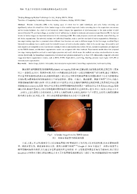
糖尿病性视网膜病变(简称糖网病,diabetic retinopathy,简称 DR)是糖尿病引起的眼部疾病,是致盲的主要原
[1]
因之一.据报道,在美国、欧洲和亚洲,大约 1/3 的糖尿病患者患有一定程度的糖网病 .研究表明,通过早期筛查,
可以显著降低糖网病的致盲率,而糖网筛查的主要方法是分析彩色眼底图像.按照国际眼科中心定义的糖网病
[1]
分级标准,与糖网病相关的眼底病变点主要包括软渗、硬渗、出血点、微动脉瘤、静脉串珠等 ,其中,出血点
和微动脉瘤的颜色偏红(如图 1 所示),统称为红色病变点.轻度非增殖性糖网病在眼底图像上的表现是只存在微
动脉瘤(microaneurysm,简称 MA),中度及重度非增殖性糖网病的表现之一是眼底存在出血点(hemorrhage,简称
HE).因此,眼底红色病变点的识别和检测对糖网病的分级诊断具有重要的指示意义.但由于病变点形态大小不
一,导致人工进行病变点分析耗时耗力,并且与大规模待检人员相比,眼科医生严重匮乏,这使得大规模的糖网
筛查难以有效开展.因此,开发一种自动化的眼底图像病变点分析工具非常必要.
Fig.1 A fundus image from the IDRiD dataset
图 1 IDRiD 数据集中的眼底图像
目前,大量工作表明,深度学习方法在眼底图像分析领域获得了比传统方法更优的性能,比如在糖网病筛
查、眼底血管分割、眼底视杯视盘分割、病变点检测等领域 [2−10] .Google 在 2016 年提出了一种基于深度学习
模型的糖网病筛查算法,首先通过多位职业眼科医生对将近 13 万张眼底图像进行病变等级标注,然后采用
inception-v3 网络对其进行训练和测试,在由 1 万多张图像组成的测试集上获得了 90.3%的敏感性和 98.1%的特