Page 297 - 《软件学报》2021年第12期
P. 297
邓成龙  等:基于随机森林的宫颈鳞癌放化疗疗效预测                                                        3961


         therapy are difficult to be effective. Therefore, it is necessary to screen patients who are not sensitive to radiotherapy and chemotherapy
         before treatment and then to explore personalized treatment plans. In view of the above problems, this paper regards the prediction of the
         efficacy  of radiotherapy and chemotherapy as  the  image classification  problem, and  proposes a model to  predict  the efficacy  of
         radiotherapy and chemotherapy for SCC based on random forests algorithm, and screens out patients who are not sensitive to radiotherapy
         and chemotherapy. First, the 3D SCC MRI (magnetic resonance imaging) is preprocessed by wavelet transform and Gaussian Laplacian;
         Second, U-net  is used  to  segment the  tumor area  in  MR  images; Then, combined with  3D  SCC MRI and corresponding tumor
         segmentation results, the texture and shape features of lesions are extracted and the extracted features are screened to train random forests.
         The experimental data set consisted of pre-treatment MR image slices of 85 patients with SCC stage IIB~IVA. The experimental results
         shows that the prediction model based on random forests predicts the efficacy of radiotherapy and chemotherapy for SCC with an AUC
         value of 0.921, which is better than the most advanced prediction method.
         Key words:    cervical squamous cell carcinoma; prediction of the efficacy of chemoradiotherapy; random forests; U-net; tumor feature
                  extraction
                                                       [1]
             宫颈癌在女性癌症疾病中发病率较高,且致死率较高 .引发女性患宫颈癌的主要因素是感染人乳头瘤病
                                                                                     [2]
         毒(human papilloma virus,简称 HPV).此外,过早的性生活、免疫抑制以及吸烟都可能引发宫颈癌 .
                                                                [3]
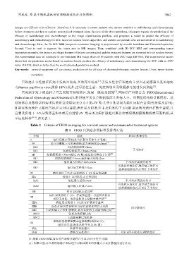
             宫颈癌多发于阴道和子宫之间的宫颈转换区,发展一般较为缓慢 .国际妇产科联合会 FIGO(International
         Federation of Gynecology and Obstetrics)妇科肿瘤学委员会根据临床上肿瘤大小、周围组织结构受累情况、远
         处转移以及影像学和病理结果将宫颈癌病变分为 I 期~IV 期.其中:I 期表现为癌灶局限在宫颈(包括累及宫体);
         II 期表现为癌灶已超出宫颈,但未到达盆壁,癌灶累及阴道,但未及阴道的下 1/3;III 期表现为癌灶扩散至盆壁,并
         且累及阴道下 1/3,导致肾盂积水或无功能肾;IV 期表现为癌扩散超出真骨盆或癌浸润膀胱黏膜或直肠粘膜,甚
         至远处转移     [4−6] ,详见表 1.
                  Table 1    Criteria of FIGO in staging for cervical cancer and recommended treatment options
                                     表 1   FIGO 宫颈癌分期标准及推荐疗法
                   分期                      描述                           FIGO 推荐疗法
                  I           病灶局限在宫颈(是否扩散至宫体不予考虑)
                                                            (1)
                   IA        仅在显微镜下可见浸润癌,最大浸润深度<5mm
                   IA1                间质浸润深度<3mm
                   IA2              间质浸润深度≥3mm,<5mm                       手术治疗
                                                              (2)
                   IB      浸润癌深度≥5mm(超过 IA 期),病灶仍局限在子宫颈
                   IB1         间质浸润深度≥5mm,病灶最大经线<2cm
                   IB2              病灶最大经线≥2cm,<4cm                   手术治疗或放疗均可
                                                                  首选同步放化疗,放疗缺乏地区可
                   IB3                病灶最大经线≥4cm
                                                                  选择新辅助化疗后再行手术治疗
                  II          病灶超出子宫,但未达阴道下 1/3 或未达盆壁
                   IIA              侵犯上 2/3 阴道,无宮旁浸润
                   IIA1                病灶最大经线<4cm                     手术治疗或放疗均可
                                                                  首选同步放化疗,放疗缺乏地区可
                   IIA2               病灶最大经线≥4cm
                                                                  选择新辅助化疗后再行手术治疗
                   IIB                有宮旁浸润,未达盆壁
                            病灶累及阴道下 1/3、扩展到盆壁、引起肾盂积水
                  III                                     (3)
                              或肾无功能、累及盆腔及主动脉旁淋巴结
                   IIIA         病灶累及阴道下 1/3,没有扩展到骨盆壁

                   IIIB      病灶扩展到骨盆壁和引起肾盂积水或肾无功能                        同步放化疗
                   IIIC   不论肿瘤大小和扩散程度,累及盆腔和主动脉旁淋巴结             (3)
                   IIIC1               仅累及盆腔淋巴结
                   IIIC2              主动脉旁淋巴结转移
                              肿瘤侵犯膀胱黏膜或直肠黏膜(活检证实)、
                  IV
                                 超出真骨盆(泡状水肿不分为 IV 期)
                   IVA                  转移至邻近器官
                   IVB                  转移至远处器官                     化疗±同步放化疗±靶向治疗
                 注:根据 FIGO 标准,如果对宫颈癌分期存在争议,应归于更早分期:
                 (1)  如果可能,可以利用影像学和病理学对临床检查的肿瘤大小及扩散程度进行补充;

